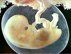
The placenta is formed as follows: a fertilized egg, entering the uterine cavity, is attached to its wall, sinking into the mucous membrane, like a "hot ball into oil." On all sides the egg is surrounded by the mucous membrane of the uterus and feeds by sweating the nutrients through the membranes of the fetal egg. After 9 days on the outer shell of the fetal egg there are villi, which penetrate the mucous membrane of the uterus, and already along them nutrients get to the fruit.
Subsequently, that part of the villi, which is facing the wall of the uterus, forms the placenta and penetrates deep into the muscular layer of the uterus. But between the villi and the wall of the uterus, there is a space in which blood circulates - here there is an exchange of oxygen, carbon dioxide, nutrients from the mother to the fetus and back.
As the pregnancy progresses, the placenta also grows. It is now more compact, dense, takes the form of a disk. One of its sides is turned towards the baby, the umbilical cord departs from the center, in which the blood vessels are located. On these vessels, nutrients, oxygen enter the fetus, and the products of its vital activity enter the mother's blood. The other side of the placenta, the mother, is attached to the wall of the uterus.
As you can see, the placenta replaces the baby with several vitally important organs: lungs, stomach, kidneys, etc. A baby can develop normally only if the placenta is working properly. The doctors of the future mother's body unite with the placenta and baby into a single system of "mother-placenta-fetus". The scale of this system is enormous, its surface is about 9 m 2 , and the network of blood vessels is 40-50 km in length! The thickness of the placenta is 3-4 cm, at the end of pregnancy its weight is 500-600 g.
The human placenta functions as a barrier, it does not let harmful substances and infectious agents pass to the baby, but, unfortunately, the chemical components of some of the medicines that mother and sometimes infectious agents can infect through it. The placenta also produces a number of hormones and other active substances that support the development of pregnancy and the growth of the baby.
The placenta has a beneficial effect on the future mother's organism, highlighting a multitude of hormones that help it to adapt to pregnancy, participate in the mechanism of the onset of labor. That is why, when watching the future mother, doctors pay special attention to the appearance and structure of the placenta during the entire pregnancy. In ultrasound examination, placental attention is paid, first of all, to the place of its attachment. Usually it is located on the bottom of the uterus or on one of its walls. But sometimes the placenta can be placed too close to the cervix. This can lead to the fact that later it will fall lower, into the area of the internal pharynx of the cervix, covering it completely (central placenta previa) or partially (marginal placenta previa).
With the development of central placenta previa, natural births are impossible - only caesarean section. This should not be frightened. In our time, the operation is performed qualitatively, without consequences for the mother and the baby. By the way, the operation may not be required. Sometimes, with an increase in pregnancy, the placenta may, on the contrary, gradually rise and occupy a normal position. Placenta prevalence threatens bleeding during pregnancy, abortion, premature birth.
In ultrasonography, placental attention is also paid to its thickness. Exceeding the permissible size can mean swelling of the placenta, which happens with Rh-conflict, diabetes, the presence of infection, malformations of the baby, severe gestosis. A decrease in size indicates placental insufficiency. In any case, it is necessary to take measures to improve the functioning of the placenta in order to ensure the normal development of the fetus. It is especially important to determine the development, maturity of the placenta in different periods of pregnancy. If the placenta begins to ripen too early, it already indicates a threat of abortion.
As soon as the baby is born, and the doctor cuts off the umbilical cord, the functions of the placenta end, and within 30 minutes the third, final phase of childbirth occurs - the birth of the placenta and membranes (afterbirth). After that, the placenta is carefully inspected - are there any defects, additional lobules, calcareous deposits (calcification), indicating that the baby in the womb suffered from insufficient nutrition. This fact must be reported to the pediatrician. After all, for a child, such information is his first health indicator or the first symptom of possible diseases. If there is a defect in the placenta, to prevent uterine bleeding, the anesthesia removes the remains of the placenta from the uterus.
So, the placenta of a person, about the structure, development, functions, you now know is a temporary but very important organ that feeds and protects the child in the womb of the mother. After birth, the placenta is either to be destroyed or used for therapeutic or scientific purposes.