Arthroscopy is a minimally invasive surgical procedure that allows visualizing the cavity of the knee joint. In addition to diagnostic tasks, some medical manipulations may be performed during arthroscopy.
Development of the method
The technique of arthroscopy was first described in 1918 in Japan. In subsequent years, the method was used only by individual specialists, and in 1957 it was brought to the attention of orthopedic surgeons all over the world. The development of medical technologies has led to a wider use of arthroscopic methods of examining the knee, ankle, hip, shoulder and wrist joints.
Advantages of arthroscopy
A significant advantage of arthroscopic surgery is that after it there is almost no scarring left. This allows you to significantly reduce the recovery period. In addition, there is no need for hospitalization of the patient after the procedure, so this intervention can be performed in a day hospital. Approximately 90% of patients with knee diseases can be diagnosed on the basis of anamnesis and clinical examination.
Magnetic resonance imaging
In some cases, patients with arthroscopy can be assigned to patients magnetic resonance imaging (MRI) or diagnostic arthroscopy. Advantages of MRI are non-invasiveness and painlessness. However, this method does not allow the simultaneous carrying out of medical manipulations.
Arthroscopy
During arthroscopy, inspection of ligaments and cartilage of the knee joint is performed. Also, the condition of the external and internal meniscus is estimated - small cartilaginous pads between the femoral and tibia.
Arthroscopy can be combined with the implementation of a number of procedures:
- treatment of meniscus ruptures (small discontinuities are usually eliminated with the help of special instruments inserted into the joint cavity);
- it is also possible to eliminate larger meniscus tears (soon after injury);
- treatment of articular cartilage damage by smoothing uneven areas with the help of special devices.
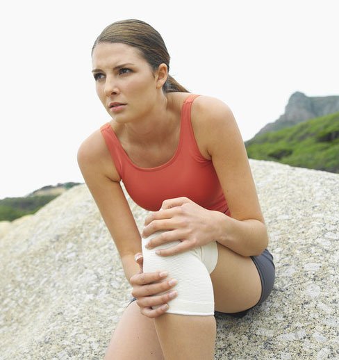
Miss Johnson, a 25-year-old professional dancer, injured her knee during the performance.
Severe pain in the knee
When the pain in the knee becomes unbearable, a woman can seek medical help. The doctor will listen to the patient's complaints and examine the knee joint. After the initial examination, it will be sent to the orthopedic surgeon of the nearest clinic for consultation and additional examination.
Specialist examination
The orthopedic doctor examined the injured knee, noting the limitation of the volume of movements - the patient could not fully bend and straighten her leg. In addition, she complained about the instability of the joint (leg in the knee as if "buckled"). The area of the joint was swollen and painful on palpation. This indicated a possible damage to the meniscus - one of the two small cartilaginous discs located in the cavity of the knee joint. The doctor suspected a rupture of the medial (internal) meniscus, possibly in combination with the anterior cruciate ligament rupture. The inner meniscus is most often damaged by a sharp turn of the shank, when the leg is bent at the knee joint.
Direction for arthroscopy
Arthroscopy of the knee joint description is prescribed by an orthopedist. To clarify the diagnosis and begin treatment of the damaged articular cartilage, the orthopedic doctor prescribed arthroscopy. The patient was admitted to the day hospital for an operation under general anesthesia. The goal of the surgical intervention was a complete restoration of the function of the knee joint. After the anesthesia began to act and the muscles surrounding the knee joint were completely relaxed, the doctor again inspected the injured limb. A repeated examination under general anesthesia often reveals a greater degree of weakening of the ligaments. A pneumatic hemostatic tourniquet is applied to the operated limb, which ensures the clamping of the vessels due to compression.
Subject to time restrictions, this procedure is safe. It greatly simplifies the process of surgical intervention. Reducing the flow of blood provides a clearer visualization of the joint cavity. To treat the operating field, the knee joint area is carefully lubricated with an antiseptic (iodine solution). The zone of surgical intervention is covered with sterile napkins. The doctor enters an arthroscope in the joint cavity, connected to the video camera. The diameter of the optical tube is 4.5 mm. The instrument is inserted from the outside of the knee joint, just below the kneecap. Using the built-in video camera, the image of the internal joint structures is transferred from the arthroscope to the monitor screen. Thus, the surgeon can examine the articular cavity and reveal the pathology of the cartilage, ligaments and menisci. The resulting image can be saved for later use.
The arthroscopic picture of the joint cavity allowed an accurate diagnosis. On the screen, the rupture of the back of the inner meniscus was clearly visible. Thus, during the arthroscopy the preliminary clinical diagnosis was confirmed. On the inner side of the joint, a second small incision (about 5 mm) is performed to insert special tools into its cavity. The damaged fragment of the cartilage is removed with the help of special tools allowing gradually, layer by layer, to "shave off" the smallest parts of it. After removing the damaged part of the meniscus, the joint cavity is thoroughly rinsed with the irrigation solution. Before closing the wound, you need to make sure that there are no particles of damaged cartilage inside. Each of the two incisions is sutured with a single stitch and sealed with a medical plaster.
After arthroscopic surgery, scarring is almost non-existent. This is one of the main advantages of this method. Places of incisions are chopped with a solution of local anesthetic, which is also injected into the joint. This allows you to minimize pain after the end of the anesthesia. Before removing the pneumatic tourniquet, an elastic bandage is applied to the knee, exerting gentle pressure on the operated area. After the termination of surgical intervention the patient was transferred to the ward for postoperative recovery. The operation did not last long. She felt slight discomfort in the knee area, but she did not feel much pain.
• Postoperative examination
After some time the patient was examined by an orthopedic doctor who reported that during the operative intervention the preliminary diagnosis of the meniscus rupture was confirmed. Before discharge, the postoperative elastic bandage was removed, and the joint was fixed with a seamless tubular bandage (elastic "stocking").
• Exercise stress
Lack of physical activity can lead to rapid muscle atrophy, so the patient needed to regularly perform a series of exercises to maintain muscle tone.
• Remote forecast
The patient was warned to avoid intensive physical exertion for at least four weeks after the operation. As the muscles of the hip are strengthened by exercise, restrictions in physical activity can be almost completely removed. The removal of a small portion of the meniscus rarely leads to complications in the future. Most patients recover completely within six weeks after surgery.