What is ultrasound used for diagnosis during pregnancy?
In the 21st century, parents do not necessarily have to wait nine months to see their baby. Thanks to modern diagnostics of ultrasound, a long-awaited meeting is possible at an early pregnancy. True, in recent years, parents increasingly do not want to know the sex of the unborn child. Thus, emphasizing the importance of birth and the girl, and the boy, and several children. However, this is not an excuse to refuse ultrasound diagnosis! Especially in the first trimester of pregnancy. What else is so useful for planned research, besides satisfying the curiosity of moms, dads and numerous relatives?
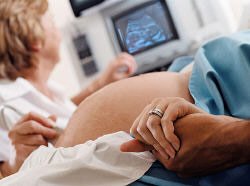
Diagnosis with the help of ultrasound in a short time became mandatory when examining every pregnant woman. Apparatus ultrasound is now in small towns, with all women's consultations. The main benefit of such studies is reliable data on the development of the fetus without causing any harm and discomfort to both. The principle of operation of ultrasound devices is simple enough: a sensor mounted on the stomach sends weak signals that, passing through the uterus, the fetus, the placenta are partially reflected and send the response signals displayed on the monitor screen. Reflected waves can be distinguished by color: dense tissue (bones) - white, soft tissue - gray, amniotic fluid - black, because for ultrasound they are transparent. On the basis of these transfers, the computer generates information according to which the doctor evaluates the child's condition and assumes its development in the future.
In conversations and discussions about the advisability of ultrasound diagnosis all the arguments "against" are suppressed by the following fact: the earlier a violation is detected in the development of the fetus, the one with the least consequences for the child and the preservation of the maternity's health can something be corrected. Alas, genetic defects and defects in children, can suddenly appear at different periods of pregnancy. And with the usual external examination of a woman, according to the results of clinical analyzes, the exact picture of what is happening is not made.
Modern methods of ultrasound
In modern medicine, various methods of ultrasound diagnosis develop every day. Examinations during pregnancy provide both physicians and parents with great opportunities to ensure the birth and appearance of healthy children. If previously the patient was examined only externally, then today you can use a vaginal sensor. This is real salvation in cases where the child is too deep or the woman is overweight.
A transvaginal long or narrow sensor is introduced in the early stages of pregnancy. It has a smaller ultrasonic power, but it increases the reliability and range of results. In addition, we used to be not always satisfied with a clear picture of the main organs and systems of the baby's body in black and white color (2D). Now parents can choose 3D or 4D diagnostics so that in dynamics, in a color image, thoroughly examine their heir. What can we say about the importance of diagnosing the blood flow of the placenta, fetal blood circulation, oxygen saturation, which became routine Doppler procedure (a kind of ultrasound).
Trying to capture every moment, starting with the joyful news from conception to the birth of a crumb, you still should not abuse modern achievements. You do not have to do ultrasounds too often just to get pictures of a child or a video with his tricks in your stomach. After all, for clarity, the doctor can increase the signal power and viewing time. Think, first of all, about the health and safety of the baby.
The terms and duration of ultrasound diagnosis during pregnancy have been officially established. The procedure is carried out for up to 30 minutes with strictly controlled signal strength and radiation range. This time is enough for both the doctor and parents. And for a picture for memory, and to ensure the safety of mom and baby. But most importantly, the doctor will reveal not only possible deviations from the norm, but also be able to predict how the pregnancy will go on.
The doctor must:
• Identify and confirm the onset of pregnancy in the early stages.
• Identify a multiple pregnancy, in order to mentally prepare parents, provide financial opportunities and tolerate babies without problems.
• Calculate the exact age of the fetus and the expected date of delivery.
• Diagnose ectopic pregnancy and in the early stages to adjust the situation without serious surgical intervention.
• To reveal the pathology of the course of pregnancy - detachment of the placenta, the threat of interruption, the tone of the uterus and other factors to keep the pregnancy.
• Identify the developmental defects of the fetus and assess their degree (incompatibility with life or the need for treatment).
• Take into account the characteristics of the birth - the mass of the fetus, the presentation, the condition of the cord, its cuff, and the date of birth.
• Find out the sex of the child.
Indications for ultrasound diagnosis during pregnancy
A solid list of information, which the doctor has after ultrasound, makes it possible not to act blindly, as in the case of a mother's refusal from such studies. Then one wrong step can do more damage than the duration of the ultrasonic radiation. And if the direction for ultrasound diagnosis is given to you in connection with the state of health, then there can be no refusal.
• Chronic diseases and various blood diseases, in which consultation of a geneticist is also mandatory.
• In the case of cases, even on the basis of the blood line of miscarriage, fetal fading, miscarriages or hereditary diseases. To do this, you will have to take an additional blood test in order to reveal the likelihood of developing a child's disease.
• If you talk about pregnancy, you worked in harmful chemical production or X-ray room.
• Suspected violations during pregnancy.
Another huge merit of ultrasound is the preservation of unwanted pregnancy. Little is said about this, but if a woman is not at first set up for change, then due to various circumstances it is decided to interrupt pregnancy. But, having heard the knock of the heart of a crumb, after seeing a real real man inside himself from the monitor, changes his opinion and gives birth!
Are you absolutely healthy?
Even absolutely healthy women doctors recommend to make ultrasound diagnosis in the first trimester of pregnancy. Having done ultrasound, you will not only be insured, but even until childbirth you will not worry about the baby. To panic, experience and experience a deep stress from a problem pregnancy is also not worth it. Modern achievements of medicine, your responsible behavior and caring for the baby, following the recommendations of the doctor will lead to a successful development of events. There are no desperate situations, and in extreme cases, many diseases are treated and adjusted in the womb.
Unreliable information about the dangers of ultrasound, designed to attract interest in certain information sources, not only does not have any research on it, but also formal reasons. Moreover, it is cruel and inhumane in its essence, because it can agitate a pregnant woman, push on the idea of refusal, make them torment themselves with questions about the health of the baby, take away precious time for correction of the subsequently revealed pathology. Be aware that ultrasound only slightly increases the temperature of the tissues and does not entail any negative consequences. From the moment of joyful news about the appearance of a child, much will depend on the personal decision of the woman and her environment. Therefore, it is better to do without extremes - for example, to refuse altogether from ultrasound or to conduct ultrasound just in case every month.
Decipher the results of ultrasound
If for some reason the results of ultrasound diagnostics cause doubts or the slightest excitement, try first to understand the unusual and unfamiliar terms yourself. After the ultrasound for the doctor of the female consultation you will be given a sheet with the data, which you can ask to decipher on the nearest reception of the gynecologist:
Fetus - the number and condition of the future baby (babies).
Preposition - head, pelvic, transverse, oblique, unstable. After 30 weeks, the fetus must occupy or already be in the head-in advance position. But if the child does not turn over by the date of birth, the cesarean section will be performed.
Fetometry of the fetus is a measurement of the head and cerebellum, abdomen, hips, tibia, heart.
The constitutional features of the structure of the fetus - the ratio of the fetal indicators for a given period and the features of the parents' constitution. Errors are allowed.
The child develops spasmodically - the definition of a possible delay in intrauterine development and the syndrome of fetal development retardation. At the slightest sign, dopplerography and cardiotocography are additionally performed. Then the child will be observed in dynamics every 2 weeks, so as not to conduct additional therapy with the risk of possible complications.
The size of the collar space is no more than 2.5 - 3 mm at the 12th week. If more, they will conduct an anamnesis, a test for alpha-fetoprotein, an examination of the blood from the umbilical cord. To exclude or confirm chromosomal diseases.
The neck implantation of the umbilical cord - determined or not, for tactics and birth management. Therefore, the indicator is absolutely not critical.
Fetal heart rate is 110 - 180 beats per minute in early pregnancy and decrease to 120-160 by the time of onset of labor.
If, after deciphering the data, there is no calming, then it's better to contact another specialist and get a sound sleep. Focus on the approved by the Ministry of Health of your country schedule of planned ultrasound, the testimony of a specialist observing you, your personal well-being. The maternal heart and intuition will not ruin, but will make maximum efforts for a healthy and happy future child!
Do an ultrasound scan on time
To hurry to do ultrasound is no longer necessary, if only because doctors do not recommend this until 10 weeks of pregnancy. Do you want to do ultrasound diagnosis before? You will only be confirmed by the fact of pregnancy and determine the number of fruits (prolific or not). Without weighty reasons, nevertheless, it is worth keeping to the planned directions, which include three mandatory ultrasound examinations : in the period of 10 to 12 weeks, at 20-24 weeks and before childbirth at 32-34 weeks. But what are characteristic of each of the periods, find out further:
The term is 5 - 8 weeks. Diagnosis: Confirmation of the fact of pregnancy. Determining the place of attachment of the fetal egg. Viability of the embryo (cardiac contractions and movement activity) . The state of the future placenta and water is assessed. Recommendations: Doctors are asked to wait longer for finding out the other nuances. If there are problems, then repeat the ultrasound can be done after 5 - 7 days.
Term is 10 to 12 weeks. Diagnosis: A statement of a progressing pregnancy. Determination of the term and the expected date of birth with an accuracy of 2 - 3 days. Measurement of the cervical region of the embryo to exclude chromosomal abnormalities Assessment of the placenta, amniotic fluid and the beginning signs of abnormalities. Recommendations: At your request, experienced professionals can already name the sex of the baby, calm down or pay attention to the existing deviations. Remind, the next very important ultrasound in 22 weeks.
The period of 20 - 24 weeks. Diagnosis: The so-called screening examination, during which the focus is on identifying or confirming the absence of malformations. Estimating the size of the fetus and the ratio with the duration of pregnancy, and even the assumption of weight at the time of delivery. Determination of the condition of the placenta, amniotic fluid.
Term is 30 - 34 weeks. Diagnosis: Evaluation of previously studied parameters, motor activity of the fetus, study of the intensity of uteroplacental blood flow with the help of a doppler.
General recommendations for 20 - 24, 30 - 34 weeks: During the examination at these times, doctors additionally assess and comment on the condition of the cervix (varies in proportion to the period of pregnancy, closed zebra, smoothing to the date of birth). If the cervix is opened prematurely, then it will be necessary to superimpose the stitching sutures. The thickness of the uterine walls is also estimated. With a seal in any part of it, you can determine the beginning tone, which can lead to the threat of termination of pregnancy. The state of the placenta (the main supplier of oxygen, nutrients and protection from external aggression) looks at the structure and degree of maturity: zero (before the 27th week), the first (from 27th to 35th), the second and the permissible third - from 32 for 36 weeks. The number and structure of water, when the main indicator is the norm of the distance of 2-8 cm between the sites of the baby and the wall of the uterus.
Immediately before birth. Diagnosis: It is done according to the testimony or desire of the mother in order to finally find out the size of the fetus, the position and condition of the child, the possible cord hanging by the umbilical cord. Recommendations: Setting up and determining the type of delivery, taking measures for emergency childbirth in the event of problems.
As we see, with ultrasound diagnosis in the first trimester of pregnancy and at later periods a very large amount of data is analyzed. And all in order to avoid trouble during pregnancy and childbirth. Therefore, ultrasound diagnosis must be done necessarily!