Planned ultrasound during pregnancy
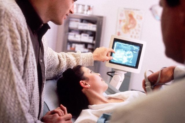
Ultrasound during pregnancy is an integral procedure that is part of the standard, planned obstetrical-gynecological follow-up of a future mother. Prinormalnom course of pregnancy, ultrasound is carried out all three times for the entire period.
The first planned ultrasound is recommended for the 10-14th week of pregnancy. It allows you to determine the exact duration of pregnancy, the position of the fetus in the uterus, the placenta state. Also, you can already detect some defects in development, reveal the signs of Down's syndrome in the fetus.
The second ultrasound is carried out on the 20th-24th week. By the time the fetus has already acquired sufficient dimensions, its heart has been completely formed, therefore it is possible to determine with greater accuracy possible defects and lag in its development, placenta previa, the number of amniotic fluid and to avoid signs of chromosomal disease. On the second planned examination, there is a high probability that you will already be told the sex of the child.
The main goal of the third ultrasound study, which is recommended for the 30th-32nd week of pregnancy, is the final assessment of the condition and position of the fetus. The doctor will determine in which presentation the baby is (in the pelvic or the head), dastotsenku his health and activity, the umbilical cord. Ultrasound at this time helps to identify such defects, which at earlier stages to identify at all was not possible.
In what cases can an unscheduled MBI be appointed?
The first so-called "ultrasound outside the plan" can be carried out at an early pregnancy with the aim of establishing the very fact of pregnancy (sometimes there is no developing pregnancy when the embryo is absent in the fetal egg) and determining its exact time, which is especially important for irregular changes.
Additional ultrasound can be performed immediately before delivery, which will predict the process of their flow.
Unplanned ultrasound examinations can be prescribed by a doctor also if a pregnant woman has some symptoms that indicate a possible pathology. The most common of these are:
- Bloody discharge from the genital tract
- Drawing pains in the lower abdomen
- Deviations in attachment of the placenta
- The mismatch of fetal size with the expected duration of pregnancy (intrauterine growth retardation)
- Threat of abortion
3D ultrasound
Today, the use of ultrasound 3D studies, also called "souvenir", is very popular. This is a relatively new research method, which allows you to see on the monitor a "photo" of an unborn child.
3D ultrasound is allowed to be performed from the 24th non-pregnancy. A three-dimensional image will give you the opportunity to get to know your little one, see his features, facial expressions and even the first smile. Such ultrasound becomes very useful for the future daddy present, since the first meeting with the baby for him is also a very important moment, especially if it is the first-born. Almost all clinics where they conduct 3D ultrasound are offered to make photos and videos with the baby. I can imagine how in a couple of years the child will be interested in looking at them.
3D ultrasound has a medical aspect of benefits: some defects (number of fingers, facial defects, nezraschivanii spinal cord, etc.) is very difficult to identify in a routine study, and 3D ultrasound offers a clearer picture, which, if necessary, allows you to change the tactics of pregnancy management. Another plural ultrasound is that the sex of the child is determined at earlier times and with greater accuracy, which is important not only to satisfy the curiosity of parents, but also in some hereditary pathologies.
Does the baby bring harm to the baby?
In fact, the opinions of specialists, and not only of our country, about the dangers of ultrasound during pregnancy diverge, because neither science nor practice has so far been able to provide us with supporting or refuting facts on this matter.
What can we say for sure? Ultrasound can deliver a child some discomfort. During this type of examination, children often turn away, begin to actively move and cover their faces with their hands, which is quite a natural reaction. They do not like very much when they are disturbed. This discomfort, as doctors say, does not carry any danger for development and health of the child.
The decision on whether to undergo ultrasound examinations only on the recommendation of a doctor or add to the initiative itself is initiative, is accepted by each parent purely subjectively and individually.
Listen to your intuition and do not neglect the recommendations of specialists. Enjoy your position!