Fertilization refers to the process of the fusion of male and female sex cells (sperm and egg), leading to the birth of a new life. Fertilization of the ovum, excreta, signs of pregnancy read on.
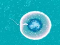
Sperm
At the end of the sexual act, the semen contained in the male seminal fluid passes through the uterine cavity. In the cervix of the uterus, sperm are fed in the alkaline medium of cervical mucus. Then they continue their movement, penetrating into the fallopian tubes (fallopian). The distance that the sperm passes is only about 20 cm, but taking into account the size of the male reproductive cell, it can take up to two hours to overcome this path.
Fight for survival
With ejaculation an average of about 300 million spermatozoa is released, but only a small part (about 10 thousand) reaches the fallopian tube where the egg is. Even less is found directly with the egg. A significant part of the spermatozoa is destroyed in the aggressive acidic environment of the vagina, and also scattered in various parts of the genital tract. Spermatozoons acquire the ability to fertilize, only after spending a certain time in the female body. The biological fluids of the genital tract activate the spermatozoa, making the undulating movements of their tails more energetic. The movement of the sperm up the genital tract is facilitated by the contractile movements of the uterus. Prostaglandins contained in seminal fluid, as well as excreted in female orgasm, stimulate these contractions.
Egg
After exiting from the follicle during ovulation, the egg is pushed out in the direction of the uterine cavity with wave-like movements of cells lining the fallopian tube. The fusion of the egg with the spermatozoon usually occurs in the outer part of the uterine tube about two hours after the sexual intercourse. On the way to the egg cell under the influence of the secret of the female genital tract, spermatozoa lose their cholesterol, which weakens their acrosomal membranes. This process is called calacitation - without it fertilization is impossible. Once near the egg, the spermatozoon is chemically "attracted" to it. Upon contact of spermatozoa with the surface of the oocyte, their acrosomal membranes are completely destroyed, and the contents of each acrosome (enzyme-containing sperm cell) leave the environment.
Penetration
Isolated sperm enzymes destroy the protective layers of the egg - cumulus mass and a shiny shell. To create a hole that is sufficient to penetrate one spermatozoon, a membrane rupture of at least 100 acres is necessary. Thus, most spermatozoa that reach the oocyte "sacrifice themselves" for the sake of introducing another sperm into its cytoplasm. After the introduction of the spermatozoon into the egg, a fusion of their genetic material takes place. The resulting zygote begins to divide, giving rise to the embryo.
Immediately after penetration of the sperm into the egg, a chemical reaction is triggered, making it impenetrable for other spermatozoa.
The second stage of meiosis
Penetration of the nucleus of the spermatozoon into the egg becomes a signal for the completion of the second reduction division (the second stage of meiosis) that began during ovulation. This forms the galloid ostida and the second polar body (which then undergoes degenerative processes). Then the nuclei of the spermatozoon and the ovum merge to form a diploid zygote that contains the genetic material of both parents.
Forming the floor
The sex of the future child is formed already at the stage of fertilization. What it will be, depends solely on the sperm. The sex of the fetus depends on the presence of the X or Y chromosome. From the mother, the fetus receives only the X chromosome, whereas from the father it can get both the X- and Y-chromosomes. Thus, if the egg is fertilized by a sperm containing an X chromosome, a female fetus develops (46, XX), and a male fetus (46, XY) when fused to a spermatozoon having a Y chromosome.
Cell division
A few hours after fertilization, a number of mitotic divisions occur in the zygote, leading to the formation of a conglomerate of cells called the morula. Morula cells divide every 12-15 hours, as a result of which it turns into a blastocyst, consisting of approximately 100 cells. Blastocyst produces a hormone called chorionic gonadotropin, which prevents the autolysis of a yellow body producing progesterone. Approximately three days after fertilization, the blastocyst begins to move along the fallopian tube into the uterine cavity. Under normal conditions, she could not overcome the sphincter of the fallopian tube. However, increased production of progesterone by the yellow body, observed after fertilization, promotes relaxation of muscles and movement of the blastocyst in the uterine cavity. Damage or overlap of the lumen of the uterine tube, which prevents the progress of the blastocyst at this stage, leads to the development of an ectopic pregnancy, in which the embryo begins to develop inside the tube.
Multiple pregnancy
In most cases, a woman has only one egg each month (alternately from each ovary). However, in some cases, eggs are excreted simultaneously from both ovaries. They can be fertilized by various spermatozoa, which leads to the development of heterozygous twins. In this case, each fetus has a separate placenta. Much less often the fertilized egg spontaneously divides into two, from which two separate embryos are formed. This leads to the development of identical twins, with an identical set of genes and a common placenta. Incomplete separation of the egg a few hours after fertilization leads to the appearance of Siamese twins.
Implantation
Having reached the cavity of the uterus, the blastocyst is implanted into the thickened mucous membrane of its wall. The hormones released by the blastocyst prevent its rejection as a foreign body. Since the successful implantation of the blastocyst, pregnancy begins.
Developmental disorders
Approximately one-third of cases of implantation of a fertilized egg do not occur, and the embryo dies. But even with successful implantation, many embryos have genetic defects (for example, an additional chromosome). Such violations often lead to the death of the embryo soon after implantation. Sometimes this occurs before the first delay in menstruation, and a woman may not even know about the pregnancy that failed.