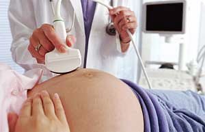
Ultrasound examinations
The first time an ultrasound is done during the first treatment of a woman to a doctor. In the early stages (5-6 weeks), the main goal of the study is to determine whether it is a pregnancy or an ectopic pregnancy. Next time, compulsory ultrasound is carried out for a period of 10 to 13 weeks. If a woman finds out that she is pregnant during this period of time, then the second planned examination becomes the first in a row. It's about ultrasound screening - a study that can identify the risk of malformations in a baby. At this stage, you can identify 2 congenital chromosomal diseases - Down syndrome and Edwards syndrome. During the next 7 days, ideally the same day, for the accuracy of the results, the expectant mother should undergo a biochemical screening, the so-called "double test". To do this, you will need to donate blood from the vein. If, based on the results of these two studies, a high risk of defects in the child is detected, the doctor will recommend prenatal diagnosis (during this procedure, amniotic fluid or cord blood is taken to analyze the chromosome set and clarify the diagnosis). The second ultrasound screening is for the 20-22nd week. Its results are also summarized with the results of biochemical screening (this time it is called the "triple test": it allows to detect also the third chromosomal disorder - neural tube defect), which is done for a period of 16 to 21 weeks. The last planned ultrasound is performed at the 32nd week. It is also aimed at detecting possible vices, undetectable due to the fact that the baby was still too small. During ultrasound, doctors evaluate various parameters that must match the duration of pregnancy: the size of the uterus and the baby, the tone of the myometrium, the degree of maturation of the placenta, the amount of amniotic fluid. Analyze the structure of the internal organs of the baby, the position of the umbilical cord.
Doppler
This method of ultrasound diagnostics makes it possible to find out whether the baby is fed enough nutrients and oxygen from the mother. During the examination, doctors assess the features of the blood flow in the uterine artery, the cord and the middle cerebral artery of the child. Having ascertained, at what speed blood flows through the vessels, it can be concluded how quickly and in what quantities nutrients and oxygen come to the baby and whether these figures correspond to the term of pregnancy. The study is conducted in 2 stages. First, each doctor examines each of the 3 arteries using an ultrasound machine. When its image appears on the screen, it turns on the sensor (Doppler), which measures the speed of the blood flow, its pressure and the resistance of the vessel. Detected blood flow disorders will indicate what complications will occur during pregnancy. So, if the baby does not have enough nutrition, he can be born with a small weight. According to the doctor's testimony, for example, if there were complications during previous pregnancies, Doppler can be performed from the 13th week. In wide practice and without fail this examination is prescribed for every pregnant woman in the period from the 22nd to the 24th week. If the doctor reveals blood flow disorders, he will prescribe a second study.
Cardiotocography
The study consists of evaluating 2 parameters - the frequency of the baby's heart rate and the state of the uterine tone. They measure 2 sensors, which are attached to the future mother on the stomach. The third one is in her hand, pressing the button every time the baby moves. The essence of the method: to analyze the change in the heartbeat of the child in response to his body movements. The goal is to find out if enough oxygen is supplied to the child. How does this method work? When we move (we run, we do gymnastics), we have a faster heartbeat. The phenomenon is called a cardiac reflex, it is formed by the 30th week of pregnancy. If we do not have enough oxygen, the heart rate will increase, and the number of beats per minute will exceed the norm. The same changes can be traced to the baby. But in the case if he is long lacking oxygen, his body will behave differently. By saving strength, the baby will move less, and in response to movement, his pulse will slow down. However, in both cases, the diagnosis is one: fetal hypoxia (lack of oxygen), only to varying degrees. As a rule, during pregnancy, the second sensor, evaluating the tone of the uterus, is rarely used. But at the time of delivery, he gives the doctor important information, showing how often the fights occur, what is their strength and duration. If they are weak, you may need to introduce drugs to enhance them. In parallel, watching the changes in the baby's heartbeat, doctors can notice and prevent other complications in time. So, if they notice that the child does not have enough oxygen, perhaps he will not be able to withstand natural births, and then he will have to do a cesarean section. KTG must be passed at least once, at the 34th week. However, many midwives advise all women to conduct this study every 10 to 14 days from the 30th week, as soon as the baby develops a cardiac reflex. The earlier the baby is diagnosed with hypoxia, the more time will remain for treatment. In some medical centers, you can rent a ktg device and conduct a study at home, sending results via video to a doctor who will monitor the situation remotely.