Every year, cases of diagnosing benign uterine tumors become more and more. Benign tumors are called differently, depending on where they develop (in what tissue). There are fibroids, fibroids, fibroids, leukemiomas.
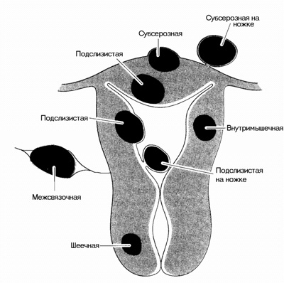
If you believe in medical statistics, the uterine fibroids diagnosed in every fifth woman, whose age is from thirty to forty-five. As a rule, these are nulliparous women. In ninety-five cases out of a hundred, the tumor develops in the uterus body and only in five cases in the cervix.
The main cause of development of a benign tumor of the uterus is an imbalance in the level of sex hormones (a decreased level of estrogen). Usually it happens during the menopause. It should be noted that a benign tumor in cancer practically does not degenerate.
Clinical picture
The clinical picture of uterine myoma is distinguished by a significant polymorphism and largely depends on the woman's age, localization, duration of the disease, the size of the formation and its morphogenetic type. In addition, the tumor can be affected by genital and extragenital comorbidities. In forty two percent of cases, the tumor for a long time does not cause any symptoms.
The probability of degeneration into a malignant tumor is very low - 0,25-0,75%, during the menopause the risk is slightly higher. However, uterine fibroids are often accompanied by pancreatic cancer, mammary glands, endometrium.
Symptoms of fibroids:
- bleeding;
- increase in the size of the tumor;
- pain;
- disruption of the work of neighboring organs.
Treatment
The appointment of a specialist with myome depends on the site of formation, the size and number of myomatous nodes, symptoms, the presence of concomitant pathology, the age of the woman and her desire to have offspring in the future, the features of the morpho- and pathogenesis of education.
Pathogenetically justified treatment of fibroids is medical and surgical, i.e. combined effect. Therefore, although many modern methods of treatment have appeared-laser, electro- and cryosurgery, the use of endoscopic techniques-treatment with hormonal preparations is also all the more urgent. The goal of conservative treatment is to reduce the severity of symptoms and (or) the size of the tumor.
The use of surgical intervention is indicated when:
- rapid increase in tumor size;
- violation of the work of neighboring bodies;
- a large amount of education (over the fourteenth week of pregnancy);
- presence of other diseases of the genital organs, which require surgical intervention;
- submucosal location of myoma, which is accompanied by abundant and prolonged menstruation, anemia;
- necrosis of the myomatous node;
- subperitoneal myoma, which has a thin base (on the "leg"); such formations are associated with a high probability of a torsion of the base of the node and with the development of its necrosis in the future;
- infertility (in those cases when it is proved that the cause of infertility is precisely this disease);
- myoma of the cervix with localization in the vagina.
Surgical intervention can be: conservative, semi-radical and radical. By the nature of access to organs located in the small pelvis, the operations can be vaginal and abdominal. The amount of intervention depends on the existing gynecological diseases (condition of the fallopian tubes, endometrium, ovaries and cervix), the age of the woman, reproductive function.
Conservative operations include:
- removal of submucous nodes;
- enucleation of nodes (otherwise, conservative myomectomy).
By semi-radical operations include:
- high amputation of the uterus;
- defundation of the uterus.
With these operations, the woman's menstruation remains, but the reproductive function is absent.
Radical operations are:
- supravaginal amputation of the uterus;
- extirpation of the uterus;
If a woman is interested in maintaining the reproductive function, then she is enucleated myomatous nodes. If the location of the tumor is sub-serous, then conservative myomectomy is performed by abdominal and laparoscopic methods. If the tumor is submucosal, the myomectomy is performed with a hysteroresectoscopy.