From the medical point of view, it is said about lop-ears when there is an angle of more than 30 ° between the auricle and the head surface or if the distance from the head to the outer edge of the ear exceeds 2 cm. In the article "Children's plastic surgery, lop-eared," you will find a lot of interesting and useful information for yourself.
Psychological problems
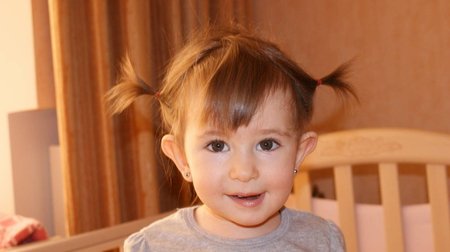
Ears are very attracting attention, and the presence of lop-earedness can give a person psychological discomfort. The idea of his body is formed in the child to about 5-7 years - that's why at this age children usually start teasing each other. Psychological trauma, received in childhood, often affects and in adulthood. There are patients who for many years dream of correcting their own shortcoming. Plastic surgery can correct this deficiency and improve the quality of life of both the child and the adult. The outer ear consists of a cartilaginous base covered with skin that provides its shape. Lop-ears can be caused by any of the following reasons (or a combination thereof):
• lack or smoothness of the antiflora. Anti-malignancy can be formed surgically. The most preferred surgical technique in such cases is modeling with incision on the front surface of the cartilage, although other methods of correction
• deep auricle. In this case, the ear looks excessively protruding. Reduction of the auricle by excision of a part of the cartilage can correct this defect;
• anterolateral rotation of the auricle in the direction from the head. Eliminate the turn can be using seams applied to the back surface of the ear and soft head tissues directly behind the ear (BTE fascia).
When surgical correction is necessary to take into account the specific features of deformity in a particular patient. Using standard operating techniques, it is impossible to achieve the desired cosmetic effect. Before the beginning of the operation, the places of the proposed incisions on the skin and cartilages of the auricle are marked with a washable surgical marker. In the space between the skin and cartilage, a local anesthetic and adrenaline are introduced to reduce bleeding during surgery, reduce pain in the postoperative period, and more easily cut the skin. After marking the place of the supposed location of the anti-curvature and the site of cartilage excision with a surgical marker, it is necessary to transfer these lines directly to the cartilage with the help of tattooing. This is necessary in order for the markings to be visible after the separation of the skin from the cartilage. Prolonged puncture of the cartilage with a needle is made along the lines on the skin. The operation begins 2-3 minutes after the introduction of a local anesthetic and epinephrine, when the drugs will begin to act. On the skin of the ear edge, silk sutures are applied (so-called suture-holders, with which one can control the position of the auricle during surgery).
The auricle is pulled forward towards the cheek, and the dumbbell outline of the skin is excised along with the underlying subcutaneous fat. This provides access to the cartilage for necessary manipulation. Since the cut is made on the back surface of the ear, after healing, the scar will be almost invisible. The cartilage of the auricle, separated from the skin, is dissected to provide access to its anterior surface. Formation of a countercut is made on the front surface of the cartilage. But in reaching the edge of the auricle, a through cut of the cartilage is made at a distance of 3 mm from the edge, along the peripheral part of the ear. The skin on the front surface of the auricle separates from the cartilage downward towards the ear canal. After dissecting the skin, cartilage of the auricle is exposed and begins to create a new shape of the ear. The sutures at this stage of the operation hold the separated skin flap outside the operating field. Each side of the cartilage of the auricle has a certain tension, which is provided by the surface layer of the cartilaginous cells and the perichondrium (a thin layer of connective tissue covering the cartilage). By making the incisions with a scalpel on the front surface of the cartilage, it is possible to attenuate the tension and deviate the auricle posteriorly, similarly to how the superficial paper layer is cut to bend the corrugated cardboard sheet. After dissecting the skin, cartilage of the auricle is exposed and begins to create a new shape of the ear.
The sutures at this stage of the operation hold the separated skin flap outside the operating field. During this procedure, an incision is made on the back surface of the auricle in the descending direction and excision of a small area of oval-shaped cartilage. To close the defect of the cartilage of the auricle, seams are applied using a resorbable suture material. It also reduces the size of the auricle and the degree of lop-ears. When tightening these seams, the auricle turns and takes a position closer to the surface of the skull. The final position of the cartilage is controlled after weakening the suture joints. At the end of the operation, you need to make sure that the bleeding is completely stopped. Otherwise, under the skin may form a hematoma (blood clots), disrupting the shape of the ear. The surgical wound and the scar must be carefully hidden behind the ear. This will hide the fact of the corrective operation. The application of the dressing is an important stage of the operation to eliminate the lop-eared. The bandage helps to form and strengthen the new position of the ear, until it finally heals. The cloth, moistened in an antiseptic, is covered with a protective layer of gauze. Then, a circular pressure bandage is applied to the head, which is fixed with a plaster to prevent it from slipping off the head or displacing. In the postoperative period, it is important to conduct adequate anesthesia. The extract is usually scheduled for the evening of the same day or the next day. After the operation, it is necessary to wear a head bandage for 10 days, then remove it. After this, the patient should apply a bandage to the auricle only on the night of the following month. Edema and bruising usually take place within two months. Patients rarely require a repeat operation, although sometimes there may be a slight deviation of the auricle from a predetermined position.