The advantages of this method are excellent quality of visualization, the possibility of obtaining images in different planes and, most importantly, the absence of any negative influence on the human body, including X-ray irradiation. This makes it possible to apply this method of diagnosis without any warning in children and pregnant women (after 12 weeks of pregnancy).
There are two types of magnetic resonance scanners: closed type and open.
A closed-type magnetic resonance tomograph is a magnetic field camera into which a person is placed for examination.
MRI of an open type has many advantages. They provide advanced imaging capabilities, a wide range of clinical applications, and an open environment during scanning. MR open-type tomographs are designed for examining patients of any age, weight, and also suffering from claustrophobia (fear of enclosed space). A C-like open type magnet provides convenient access to the patient during the diagnostic procedure, allowing the family member or doctor to be in close proximity to a small child, severely ill or a patient of advanced age. A large viewing angle increases the comfort of the patient being examined, minimizes claustrophobia and anxiety during the procedure.
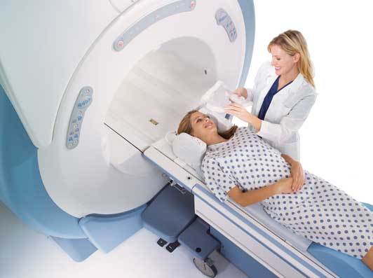
How is the MRI examination performed?
On average, the duration of the diagnostic procedure of magnetic resonance imaging ranges from 30 to 60 minutes, during which the magnetic field generates radio waves that are sent to specific areas of the body. Received from the monitored organs echoes, the computer program transforms into layered images. In this way, pathological changes in the body (eg, prolapse of the disc, breast cancer or brain pathology) can be reliably diagnosed without the use of X-rays. During the diagnostic procedure, it is advisable to lie still and breathe evenly. The slightest movement can cause distortion of the image, and accordingly, and limit the accuracy of the diagnosis.
During the magnetic resonance imaging, the patient does not experience any pain sensations, except for a feeling of light heat in the part of the body being examined.
Indications for magnetic resonance imaging.
MRI diagnostics is performed exclusively on indications in the presence of a referral indicating the area of study and the diagnosis of the doctor, the clinical situation or the purpose of the diagnosis.
Indications for MRI of the head:
- Anomalies and malformations of the brain.
- Post-traumatic injury.
- Inflammatory processes and infectious diseases.
- Multiple sclerosis.
- Vascular disorders (strokes, hematomas, aneurysms, malformations).
- Tumors of the brain and its membranes.
Indications for MRI of the spine and spinal cord:
- Injuries of the spine.
- Hernia of intervertebral discs.
- Inflammatory processes of the spine and spinal cord.
- Vascular disorders (strokes, hemorrhages).
- Tumors of the spinal cord and spine.
- Scoliosis.
- Congenital diseases.
- Degenerative and dystrophic processes.
Indications for MRI of the musculoskeletal system:
- Traumatic injuries of bones, muscles, ligamentous apparatus.
- The defeat of the meniscus.
- Osteonecrosis.
- Inflammatory processes of bone tissue (tuberculosis, osteomyelitis).
- Degenerative and dystrophic processes.
- Tumors of bones and muscles.
- Diseases of bone marrow.
Indications for MRI of the chest and mediastinum:
- Vascular anomalies.
- Anomalies, malformations of the tracheobronchial tree.
- Tumors of the mediastinum.
- Hematological diseases.
- Myasthenia gravis.
- Injuries, inflammatory processes, tumors of the soft tissues of the chest.
Indications for MRI of the abdominal cavity and retroperitoneum:
- Tumors of the parenchymal organs (liver).
- Retroperitoneal fibrosis.
- Lesions of the spleen, lymph nodes in hematological diseases.
- Visualization of prevalence of aortic aneurysm.
Indications for MRI of the pelvic organs:
- Tumors of the genital organs.
- Tumors of the urinary system, rectum.
- Endometriosis.
- Inflammatory processes, fistulas.
- Anomalies, malformations of the pelvic organs.
How to prepare for the MRI procedure?
Since a strong magnetic field inside the device will attract any object that contains iron or some other magnetic metal, the doctor who will conduct the research should ask if you do not have metal implants (for example, hip prostheses, heart valves, pacemakers , as well as bullets, fragments, etc.). The same applies to bras with metal hooks-fasteners, zippers, buttons and other metal parts on clothes - they complicate the adjustment of the device, and sometimes distort the image, which complicates the diagnosis. The doctor will ask you to remove such clothes, as well as ornaments (rings, earrings, chains, watches), change into a disposable gown and change shoes.
Dental fillings, crowns, bridges, as a rule, allow to conduct a survey, although metallic oral implants affect the magnetic field, which worsens the image of the mouth area.
A strong magnetic field can irretrievably damage mobile phones, electronic devices (hearing aids, pacemakers) wristwatches, storage media (including credit cards). For the duration of the examination, it is necessary to leave such items in a personal closet or to deposit it with a doctor.
During the MRI of the head, makeup elements (mascara, shadow, powder) can interfere with obtaining quality images and reduce their diagnostic value. Going to the MRI diagnosis, women are advised to refrain from applying make-up or to remove go immediately before the procedure.
If you read these lines long before the examination, then, going to the MRI diagnosis, try to dress accordingly.
Special preparation for MRI is not required. You can eat, drink, take medicine in the usual way for you. If you need special training, with some studies on MRI, you must be warned in advance.
If you have ever felt panic or fear in a confined space and you have to be examined on a magnetic resonance tomograph of a closed type, then inform the doctor about it.
As a rule, the examination is not carried out in the first 12 weeks of pregnancy, with the exception of extreme necessity in the presence of vital indications or with suspicions of an abnormality in the fetus.
Children under five years for the diagnostic procedure may need a shallow general anesthesia. This should be discussed with an anesthesiologist in advance. The cost of anesthesia or contrast agent, which is used to visualize blood vessels, is usually not included in the cost of the MRI procedure itself and is paid for separately.
Be patient while going to the MRI diagnosis - sometimes it can happen that you have to wait. Patients who are in urgent medical interventions can save lives or significantly improve the outcome of treatment are taken out of turn. Remember that somebody might be in their place, and also that there are always those who are much worse than you. Therefore, plan your affairs so that you have several hours left. And be healthy!