Shares of the brain
The large hemispheres are subdivided into parts whose names are given by the bones covering them: • The frontal lobes are located in front of the Roland and over the Sylvian furrow.
• The temporal lobe lies behind the central and above the posterior portion of the lateral sulcus; it extends back to the parieto-occipital furrow - a gap separating the parietal lobe from the occipital, which forms the posterior part of the brain.
• The temporal lobe is the area located under the sylvian furrow and bordering from behind with the occipital lobe.
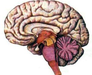
As the brain intensively grows before birth, the cerebral cortex begins to increase its surface, forming folds, which leads to the formation of a characteristic appearance of the brain resembling a walnut. These folds are known as convolutions, the grooves dividing their grooves are called furrows. Certain grooves in all people are located in the same place, so they are used as guidelines for dividing the brain into four parts.
Development of convolutions and furrows
Furrows and convolutions begin to appear on the 3-4th month of development of the fetus. Until then, the surface of the brain remains smooth, like the brain of birds or amphibians. The formation of a folded structure provides an increase in the surface area of the cerebral cortex in conditions of a limited volume of the cranium. Different parts of the cortex perform specific, highly specialized functions. The cerebral cortex can be divided into the following areas:
• Motor zones - initiate and control body movements. The primary motor zone controls the arbitrary movements of the opposite side of the body. Directly in front of the motor cortex is the so-called premotor cortex, and the third region - an additional motor zone - lies on the inner surface of the frontal lobe.
• Sensory areas of the cerebral cortex perceive and generalize information from sensitive receptors throughout the body. Primary somatosensory zone receives information from the opposite side of the body in the form of impulses from sensitive receptors of touch, pain, temperature and position of joints and muscles (proprioceptive receptors).
The surface of the human body has its "representations" in the sensory and motor sites of the cerebral cortex, which are organized in a certain way. Canadian neurosurgeon Wilder Penfield, who practiced in the 1950s, created a unique map of the sensory areas of the cerebral cortex, which perceive information from various parts of the body. As part of his research, he conducted experiments in which he suggested that a person under local anesthesia describe his feelings at a time when he stimulated certain areas of the surface of the brain. Penfield found that stimulation of the postcentral gyrus caused tactile sensations in specific areas on the opposite half of the body. Other studies have shown that the volume of motor cortex responsible for different areas of the human body depends more on the level of complexity and accuracy of the performed movements than on the strength and volume of muscle mass. The cerebral cortex consists of two main layers: the gray matter is a thin layer of nerve and glial cells about 2 mm thick and a white substance that is formed by nerve fibers (axons) and glial cells.
The surface of the large hemispheres is covered with a layer of gray matter, the thickness of which varies from 2 to 4 mm in different parts of the brain. The gray matter is formed by the bodies of nerve cells (neurons) and glial cells performing a supporting function. In most of the cerebral cortex, six separate layers of cells can be detected under a microscope.
Neurons of the cerebral cortex
The bodies (containing the cell nucleus) of the neurons of the cerebral cortex differ substantially in their form, nevertheless, only two major ones are distinguished.
- Pyramidal cells derive their name from the shape of the body of the neuron, which resembles a pyramid; their axons (nerve fibers) emerge from the cerebral cortex and carry information to other parts of the brain.
- Non-pyramidal cells (all the others) are designed to perceive and process information from other sources.
The thickness of the six layers of cells that form the cerebral cortex varies greatly depending on the area of the brain. The German neurologist Corbinian Broadman (1868-191) investigated these differences by staining the nerve cells and viewing them under a microscope. The result of Brodmann's scientific research was the division of the cerebral cortex into 50 separate sites on the basis of certain anatomical criteria. Subsequent studies have shown that the "Brodmann fields" thus isolated play a specific physiological role and have unique ways of interacting.