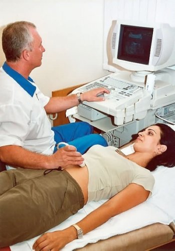
Currently, ultrasound, or in another way, echography, is the safest, most common and highly effective way of assessing the course of pregnancy. Transvaginal echography allows you to see a fetal egg located in the uterine cavity 21 days after conception, and after four - and the inhabitant of the egg.
It is important that the first during the pregnancy, ultrasound carried a specialist who is well versed in early prenatal diagnosis. Then he will be able to make sure that the child is all right, will be able to help future parents look at the ears, eyes and pens of the future baby.
All pregnant women are concerned about the safety of ultrasound. In 1978 (then ultrasound was not massively applied), studies were carried out of the biological aspects of the influence of ultrasound on living tissues. The results indicate that even in the case of multiple excess of the standard intensity of ultrasound, no harm is caused to the mammalian embryos during the diagnostic process.
In the first trimester of pregnancy, the rejection of ultrasound carries a negative effect. Each year, according to statistics, about one hundred children in Moscow are born with Down syndrome. Not all doctors know that it is possible to suspect this serious disease with the help of echography at a period of 12-13 weeks. It is very important to conduct the first ultrasound examination before the end of this period. Let's see why.
- During this period, the best malformations of the fetus and the markers of chromosomal pathology are best diagnosed. Just a few weeks later, features of ultrasound that can recognize Down's syndrome and other serious diseases can disappear without a trace.
- In case of suspicion of chromosomal pathology, doctors have time to conduct a special genetic study and in case of an adverse result to terminate the pregnancy.
- The first trimester is optimal for the establishment of gestational age of the fetus with an accuracy of several days. In the event of any difficulties, obstetricians will be guided precisely at this age.
In the early stages of pregnancy, ultrasound is done in pursuit of such goals:
- Proof of pregnancy. Ultrasound is the most accurate method of detecting pregnancy in the early stages.
- Definition of the term. At the fifth week with the help of ultrasound, you can accurately determine the duration of pregnancy. The error is only 2-3 days, because at this time all the fruits are almost the same.
- The location of the egg. In the early stages of pregnancy, an ectopic pregnancy can be detected using ultrasound. Sometimes there are cases when there is a localization of 2 fetal bags at once. In this case, one is located correctly, and the other is outside the uterus. To check whether there is an ectopic pregnancy, the first ultrasound is performed most often after 10 days of delay.
- Vitality of the embryo. Ultrasound in the fifth week of pregnancy allows you to fix the movement of the fetus, and in the fourth week you can see how the heart of the embryo is shortened.
- The number of fruits. At a period of six weeks, ultrasound can detect the presence of two yolk and fruity sacks. With the development of two fetuses in the uterus, the risks increase significantly (approximately 10 times). In this case, several ultrasound can not do, it is necessary to do it more often.
- Imitation of pregnancy. In pregnancy, thanks to the first ultrasound, you can find out whether imitation of conception by ovarian cysts, uterine fibroids or some types of tumors occurs.
- Threat of abortion. If a woman has spotting at the initial stages, it is necessary to know whether the heart of the embryo works correctly. At 3-4 weeks, ultrasound can be used to determine if placental abruption does not occur, and if so, then monitor the degree of detachment in order to reduce the probability of miscarriage to a minimum.
During pregnancy, the first ultrasound in the early stages can help determine the condition of the placenta. All further ultrasound procedures provide an opportunity to more accurately determine how pregnancy proceeds.