Symptoms of cataracts
In cataracts, the passage of light rays through the eye is impaired. Small cataracts may not cause any noticeable symptoms. Larger ones can be the cause of the following changes: a decrease in visual acuity ("fog in the eyes") - violates the usual actions, such as reading or driving a car; vision is often worse in bright light, and remote and central; spots - can be observed at a fixed location in the field of view; Diplopia (double vision) can be observed only on one eye and persist when the second eye is closed; glaucomatous halos - orange rings visible by the patient around light sources or any bright objects, everything around has a light orange tint; easier reading - patients who previously needed glasses for reading, sometimes do not use them anymore. Cataract-related changes in lens shape increase myopia.
Causes
Lens clouding can be: age-related - degenerative processes develop in the lens; congenital - due to an intrauterine viral infection, such as rubella, or metabolic disorders such as galactosemia, accompanied by an elevated level of galactose in the blood; hereditary - in some families there is a genetic predisposition to the development of cataracts at an early age; traumatic - due to bruises of the eye, penetrating wounds of glass fragments or metal fragments, or previous eye operations; inflammatory - patients with chronic iris of the eye (iritom) are at increased risk; caused by diabetes - with a high level of sugar in the blood, the lens can be damaged; radiation - with prolonged exposure to sunlight or ionizing radiation; caused by corticosteroids - prolonged use of drugs of this group can cause cataracts; related to skin diseases, such as atypical dermatitis. Diabetics who use insulin can also suffer from cataracts due to impaired nutrition of the lens of the eye.
Diagnostics
The diagnosis of cataracts is made after a complete examination of the eye in order to exclude other pathologies, for example glaucoma or retinal disease. Patients with cataracts are able to indicate the location of the light source, their pupils normally react to light. In advanced cases, the lens may appear brown or white.
Ophthalmoscopy
Using an ophthalmoscope (a special tool for the internal examination of the eye), one can confirm the presence of cataracts. When a ray of light is passed through the pupil from a distance of approximately 60 cm, the posterior wall of the eye normally looks red (hence the "red eyes" that are visible in some photographs). Cataract is seen as a dark spot.
Congenital cataract
All newborns, as well as children between the ages of 6 and 8 weeks should be screened for cataract and other eye diseases. Congenital cataracts must be treated within the first three months of life. In the absence of timely treatment, a development of normal vision may be disrupted, even if cataract at a later age is removed. Ophthalmologists use an ophthalmoscope for an internal examination of the eye, with the help of which it is possible to confirm or exclude the diagnosis of cataract. There is no medical treatment for cataracts. In the early stages, dark glasses can prevent eye irritation when exposed to bright light. Good lighting from the top and the back can help with reading.
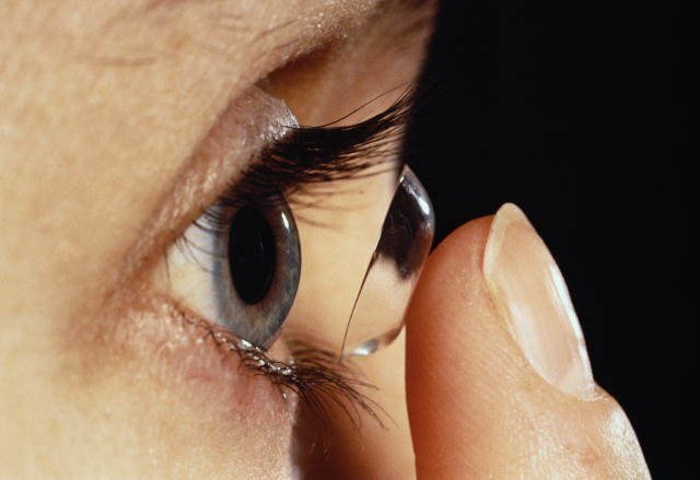
Operative treatment
The operation to remove cataracts (extraction of cataracts) is safe and effective. This is the most common planned operation in the elderly. In Russia, more than 300 thousand cataract extractions are performed annually. Among the patients, it is believed that cataract extraction is recommended only at a late stage, with significant visual impairment. With the use of modern surgical techniques, delay in the operation is not required. In extracapsular cataract extraction, the central, denser part of the lens (nucleus) can be diluted before removal by ultrasound. After surgery, most patients notice a significant improvement in vision. However, reading may still require points. The operation is usually performed under local anesthesia, with a one-day hospitalization.
Surgical techniques
Extracapsular extraction is most widely used. Using a microsurgical technique, the doctor removes the lens through a small incision of his capsule. Intracapsular extraction consists in removal of the whole lens together with the capsule, usually with the aid of a cryoprobe; this technique is currently used in a limited way. Patients usually recover quickly. In some cases, the use of anti-inflammatory and antibacterial eye drops is required for several weeks. Without the lens, the eye sees at a distant distance, but can not focus on nearby objects. Glasses or implantation of an artificial lens help to correct vision. Glasses - necessary after the operation, they increase the nearby objects, but are cumbersome and limit the field of view; the use of intraocular implants avoids the use of glasses. Intraocular implants - the development of intraocular lenses (artificial lenses) have been carried out since the Second World War, when it was discovered that fragments of plexiglas from aircraft cabs, lingering in the eye, do not do him harm, unlike many other foreign bodies. Most implantable artificial lenses are now placed in an empty lens capsule. There are various types of artificial lenses, including rigid polymethyl-methacrylate and flexible silicone lenses, introduced through a minimal incision. Cataract tends to grow over time and can subsequently cause blindness. By inhibiting the medical examination of the inside of the eye, it worsens the diagnosis of other curable eye diseases. The operation restores normal vision in the absence of another eye pathology. During the corrective operation with cataracts a cut is made along the edge of the cornea (the area is circled by a circle). This allows the wound to heal without stitching. After lens implantation, a capsule thickening is sometimes observed, which causes progressive deterioration of vision. In this case, laser treatment may be required. Cataract is a common cause of vision impairment in the elderly.