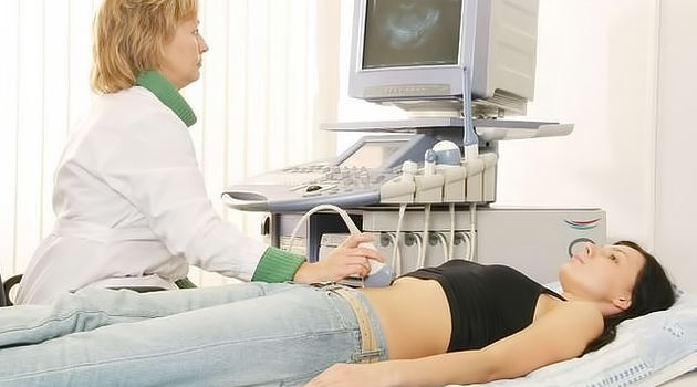
Ultrasound examination (sonography, ultrasound tomography, synovial ultrasound, ultrasonography) is currently one of the most well-known methods of medical imaging around the globe. This technique has earned its popularity due to its rich capabilities in diagnosing a wide variety of thyroid gland diseases, cardiovascular system, evaluation of fetal development in pregnancy, kidney disease, abdominal cavity organs, breast diseases. As for gynecology, ultrasound of pelvic organs in women is an important diagnostic tool in identifying problems with these organs.
At the moment, ultrasonic diagnostics have been used for almost half a century. During this time, it has passed more than one stage of development, from the moment when its results were almost not believed, until the period when its possibilities were evaluated for the dignity and ubiquity of this method. Today it is hardly possible to imagine medicine without the use of ultrasound diagnosis.
The ultrasonic method of tomography is based on the same principle as the echo sounders, that is, on the phenomenon of reflection of an ultrasonic wave from the viscera of the body. Reflected waves are captured by a special sensor, after which, based on the readings of this sensor, a planar image of the tissues and organs through which the wave passes is constructed.
On which day of the cycle is it necessary to conduct ultrasound?
If it is necessary to diagnose the presence of various formations in the small pelvis, such as ovarian cyst, uterine fibroids, ovarian fibroids and others, the day of the menstrual cycle does not matter for the passage of ultrasound, especially if the doctor is highly qualified.
In some cases, in order to successfully carry out a differential diagnosis, you may need dynamic ultrasound control, that is, you will need to perform several ultrasound examinations at various days appointed by the doctor.
Dynamic control is also necessary during the stimulation procedure to control the growth of the endometrium and follicles, as well as when registering ovulation. The most relevant is in cases where there is a pathology of the endometrium (hyperplasia, polyps) or functional ovarian cysts. In these cases, the diagnosis can be made only after several procedures of ultrasound.
Types of ultrasound
There are three types of ultrasound:
- Transabdominal examination. With it, the examination is carried out through the abdominal front wall. With this kind of research, it is necessary that the bladder is complete - thanks to this, you can clearly see the necessary organs. Such a study is carried out mainly only in the diagnosis of the abdominal cavity organs and formations in the small pelvis.
- Vaginal examination. With him, as can be understood from the name, the sensor is inserted into the patient's vagina. At this type of examination, it is necessary that the bladder is empty. Basically this type is used with a careful examination of organs located in the pelvic area.
- Transrectal. In this case, the sensor is placed in the rectum. This type of research is used in cases where the girl is a virgin, or in men in the diagnosis of the condition of organs and tissues of the pelvis.
There is Doppler ultrasound, it is necessary in the diagnosis of blood supply problems in the tissues and organs under investigation.
What can be seen with ultrasound of pelvic organs in women?
If the ultrasound procedure is performed correctly, you can see:
- The cervix and the body of the uterus;
- The rectum;
- Bladder;
- Ovaries;
- Fallopian tubes (only if there is a pathology).
The timing and indications for the use of ultrasound in the pelvic area are determined primarily by the doctor who examines you. It should be remembered that most reproductive system diseases in women can not manifest themselves at all, especially at the initial stages of their development, therefore it is recommended to undergo this examination at least once a year.
In conclusion, it can be said that at the moment, ultrasound tomography of the pelvic organs is one of the most informative, affordable, safe and economical methods of diagnosing female health.