Chromosomes contain genetic information in the form of genes. The nucleus of each human cell, with the exception of the egg and sperm, contains 46 chromosomes, forming 23 pairs. One chromosome in each pair is derived from the mother, and the other from the father. In both sexes, 22 out of 23 pairs of chromosomes are the same, only the remaining pair of sex chromosomes is different. Women have two X-chromosomes (XX), and in men there is one X- and one Y-chromosome (XY). Consequently, the normal set of chromosomes (karyotype) of the male is 46, XY, and the female - 46, XX.
Chromosomal abnormalities
If the error occurs during a particular type of cell division, in which oocytes and spermatozoa are formed, then anomalous germ cells arise, which leads to the birth of offspring with chromosomal pathology. Chromosomal imbalance can be both quantitative and structural.
Development of a child's sex
Under normal conditions, the presence of the Y-chromosome leads to the development of the male fetus, regardless of the number of X-chromosomes, and the absence of the Y-chromosome - to the development of the female fetus. Anomalies of sex chromosomes have a less destructive effect on the physical characteristics of the individual (phenotype) than the anomalies of the autosomal ones. Y-chromosome contains a small number of genes, so its extra copies have minimal impact. Both men and women require the presence of only one active X chromosome. Excess X-chromosomes are almost always completely inactive. This mechanism minimizes the effect of abnormal X-chromosomes, since superfluous and structurally abnormal copies are inactivated, leaving only one normal X chromosome "working". However, there are some genes on the X chromosome that avoid inactivation. It is believed that the presence of one or more two copies of such genes is the cause of abnormal phenotypes associated with imbalance of sex chromosomes. In the laboratory, chromosome analysis is carried out under a light microscope at a 1000-fold magnification. Chromosomes become visible only when the cell is divided into two genetically identical daughter cells. To obtain chromosomes, blood cells are used which are cultured in a special medium rich in nutrients. At a certain stage of division, the cells are treated with a solution that causes them to swell, which is accompanied by "unraveling" and separating the chromosomes. The cells are then placed on a microscope slide. As they dry up, the cell membrane ruptures with the release of chromosomes into the external environment. Chromosomes are colored in such a way that on each of them appeared light and dark discs (strips), the order of which is specific for each pair. The shape of the chromosomes and the nature of the discs are carefully studied in order to identify each chromosome and identify possible anomalies. Quantitative anomalies occur when there is a lack or excess of chromosomes. Some syndromes that develop as a result of such defects have obvious signs; others are almost invisible.
There are four main quantitative chromosomal abnormalities, each of which is associated with a certain syndrome: 45, X - Turner syndrome. 45, X, or the absence of a second sex chromosome, is the most common karyotype in Turner's syndrome. Individuals with this syndrome have a female gender; often the disease is diagnosed at birth due to such characteristic features as skin folds on the back of the neck, swelling of the hands and feet and low body weight. Other symptoms include short stature, a short neck with pterygoid folds, a broad chest with widely located nipples, heart defects and abnormal forearm deflection. Most women with Turner's syndrome are sterile, they do not have menstruation and do not develop secondary sexual characteristics, particularly the mammary glands. Virtually all patients, however, have a normal level of mental development. The incidence of Turner syndrome is between 1: 5000 and 1:10 000 women.
■ 47, XXX - trisomy of the X chromosome.
Approximately 1 in 1000 women has karyotype 47, XXX. Women with this syndrome are usually tall and thin, without any obvious physical abnormalities. However, they often have a decrease in the intelligence factor with certain problems in learning and behavior. Most women with trisomy X-chromosomes are fertile and are able to have children with a normal set of chromosomes. The syndrome is rarely detected due to the blurred manifestation of phenotypic traits.
■ 47, XXY - Klinefelter's syndrome. Approximately 1 in 1,000 men have Klinefelter syndrome. Men with a karyotype of 47, XXY look normal at birth and in early childhood, with the exception of small problems in learning and behavior. Characteristic signs become noticeable during puberty and include high growth, small testicles, lack of spermatozoa, and sometimes insufficient development of secondary sexual characteristics with enlarged mammary glands.
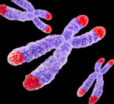
■ 47, XYY - XYY syndrome. An additional Y chromosome is present in about 1 in 1,000 men. Most men with XYY syndrome look normal, but they have very high growth and a low level of intelligence. Chromosomes in shape remotely resemble the letter X and have two short and two long arms. Typical for Turner syndrome are the following anomalies: an isochromosome on the long arm. During the formation of eggs or spermatozoa, the separation of chromosomes occurs, in violation of the divergence of which a chromosome with two long shoulders and a complete absence of short chromosomes may appear; ring chromosome. It is formed due to the loss of the ends of the short and long arms of the X chromosome and the connection of the remaining sections to the ring; deletion (loss) of a part of the short arm by one of the X chromosomes. Anomalies of the long arm of the X chromosome usually cause dysfunction of the reproductive system, for example premature menopause.
Y-chromosome
The gene responsible for the development of the masculine embryo is located on the short arm of the Y chromosome. Deletion of the short arm leads to the formation of a female phenotype, often with some signs of Turner's syndrome. Genes on the long shoulder are responsible for fertility, so any deletions here can be accompanied by male infertility.